


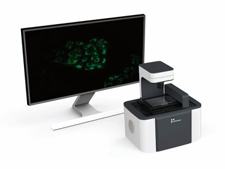
UltraView Digital Cell Imager can be placed in a Co2 incubator, that is used for observation and quantification of cell behavior via real-time, quantitative live-cell analysis. The application provides user-friendly control of all aspects of imaging and analysis including powerful and intuitive image processing tools.
Features
1. User friendly, operation simply and freely
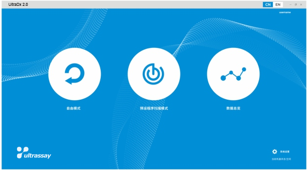
2. Intelligent automation
3. Low power consumption, high throughput
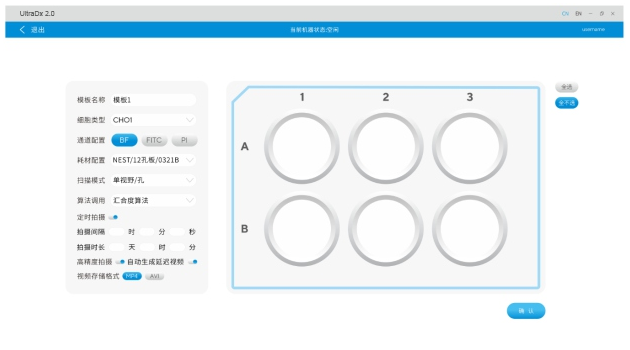
The first step is to select the vessel in the database, including 6-384 well plate.
Cell culture plates do not fog because of low power consumption.
4. Clearer images
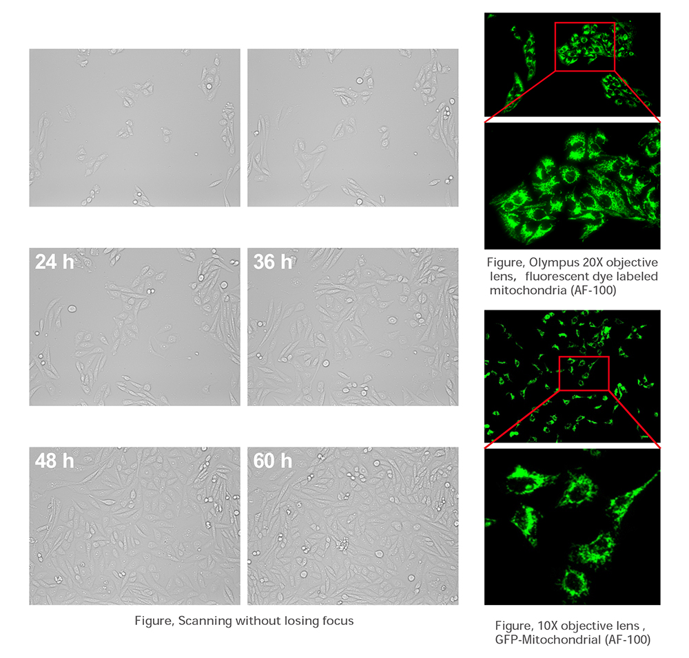
UltraView supports 4 x, 10 x, 20 x objective lens, with CMOS highly sensitive camera.
UltraView uses real-time digital phase difference algorithm to overcome low contrast and unclear imaging.
5. Auto-imaging
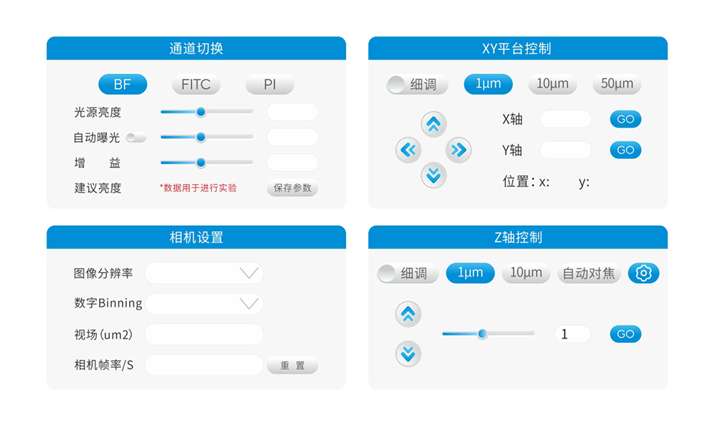
UltraView is equipped with a four-axis fully automatic switching system, which allows free switching between bright field and dual fluorescence channels. UltraView has both auto focus and manual focus and supports programmed scanning imaging.
6. Intelligent algorithm
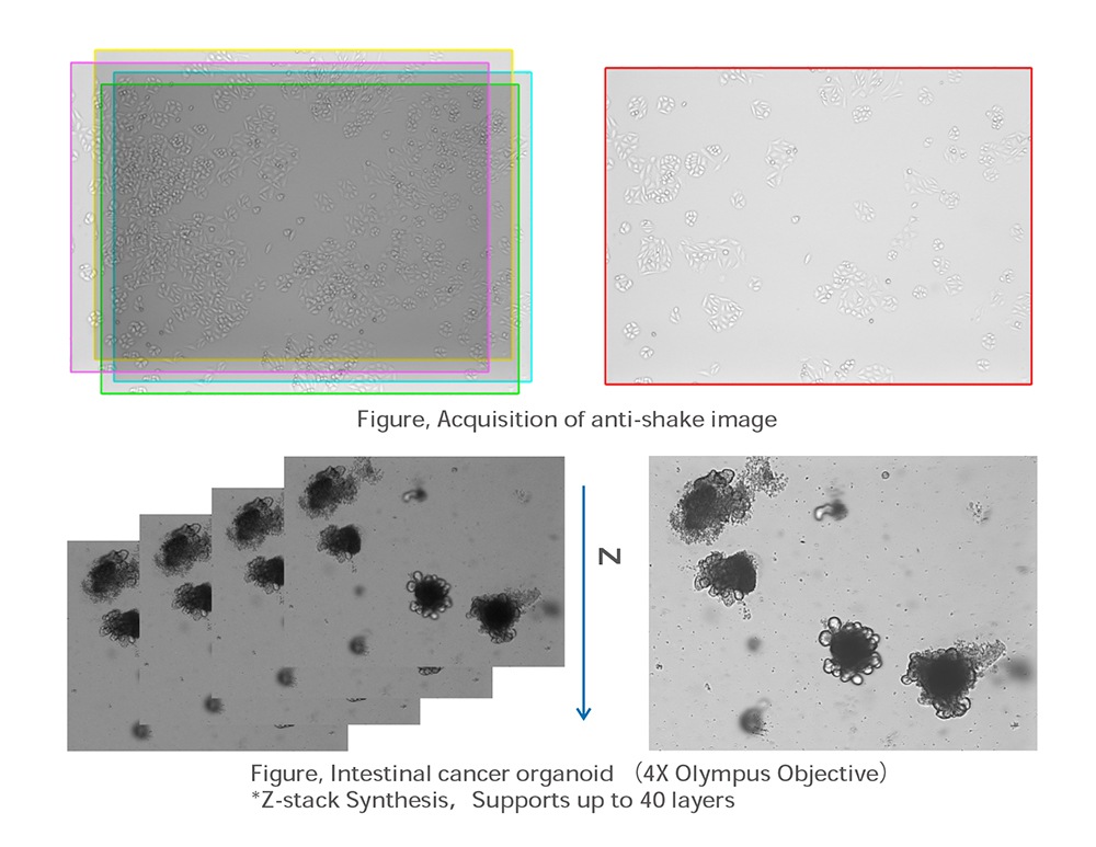
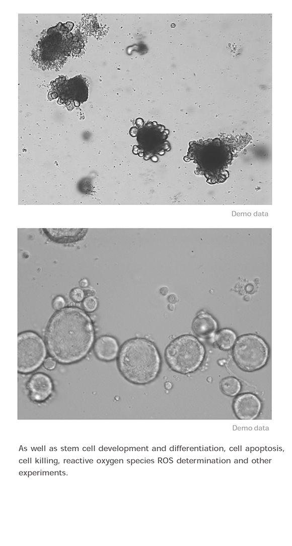
UltraView automatically creates retrospective video and supports layer scanning for tumor spheres and organoids.
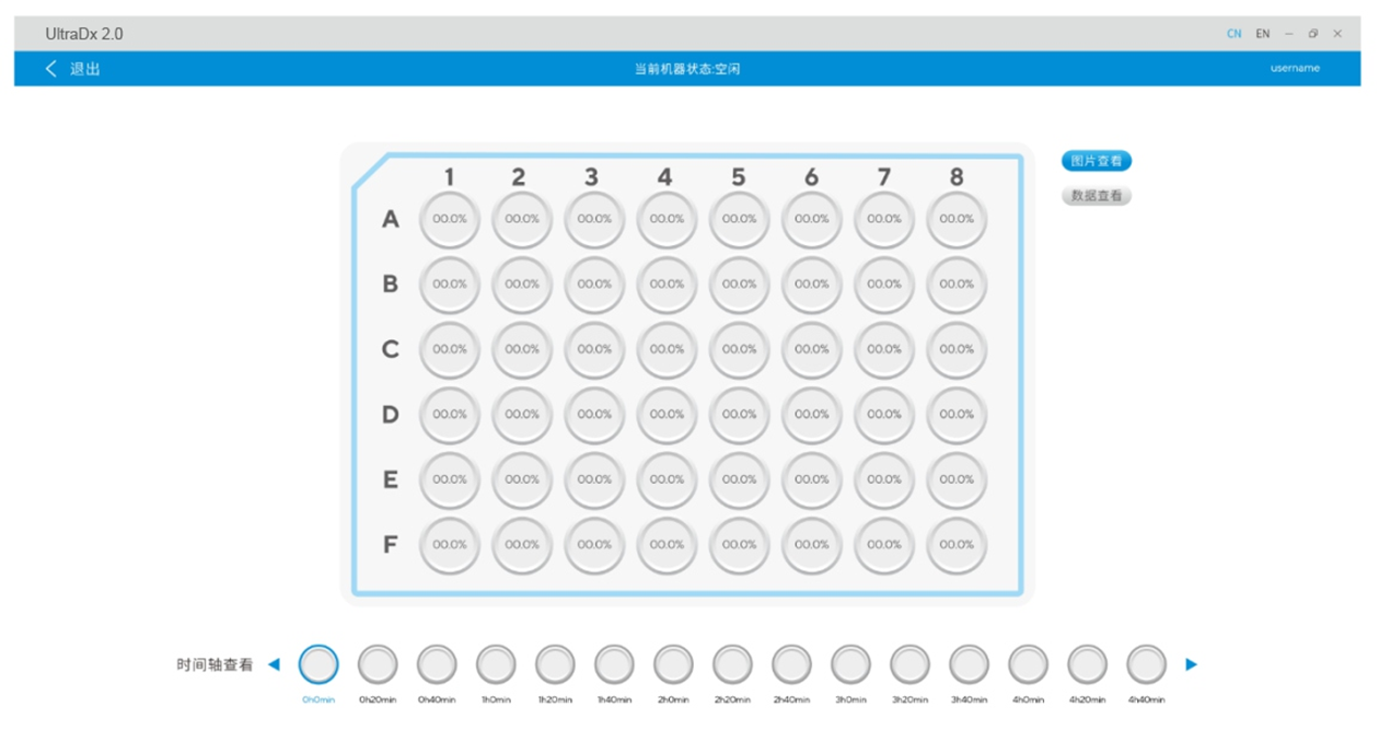
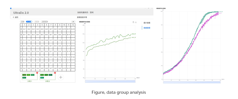
7. Data visible
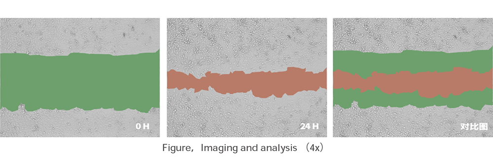
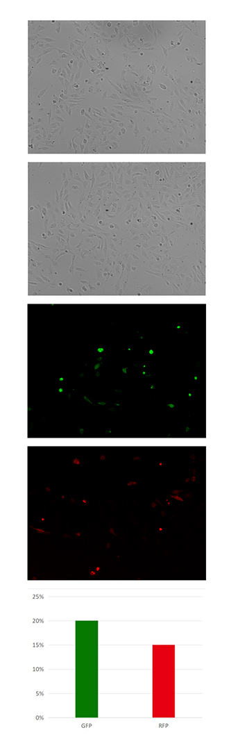
Matrix data overview, support data grouping analysis, automatic generation of proliferation curves, automatic display of cell confluence, wound healing data analysis, transfection efficiency analysis, etc.
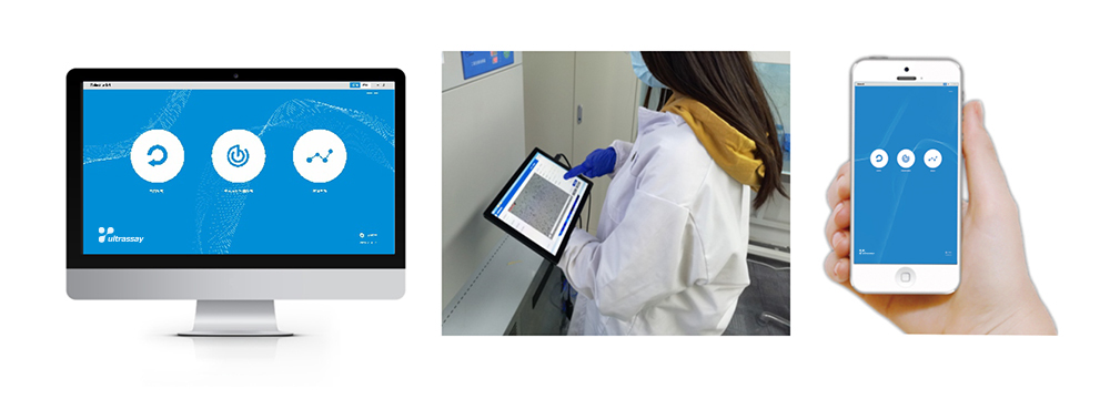
8. Three-end interconnection based on web architecture
The equipment is equipped with high performance host, LAN can support mobile phones, tablet remote Settings and view the shooting data.
9. Support customized research and development
Support customized z-stack layer scan, customized data analysis mode, etc.
Application
The instrument is widely used with multi-channel fluorescence imaging, multi-site well plate scanning, digital phase, real-time imaging and visualization of proliferation curves, etc.
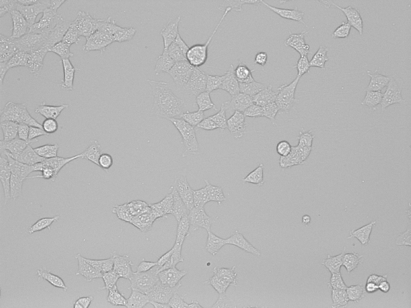
1. Bright field live cell real-time marker-free imaging
It can monitor cell growth and cell culture quality, optimize culture conditions (such as media development, etc.).
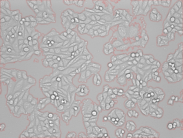
2. Cell proliferation and confluency analysis
3. Wound healing assay
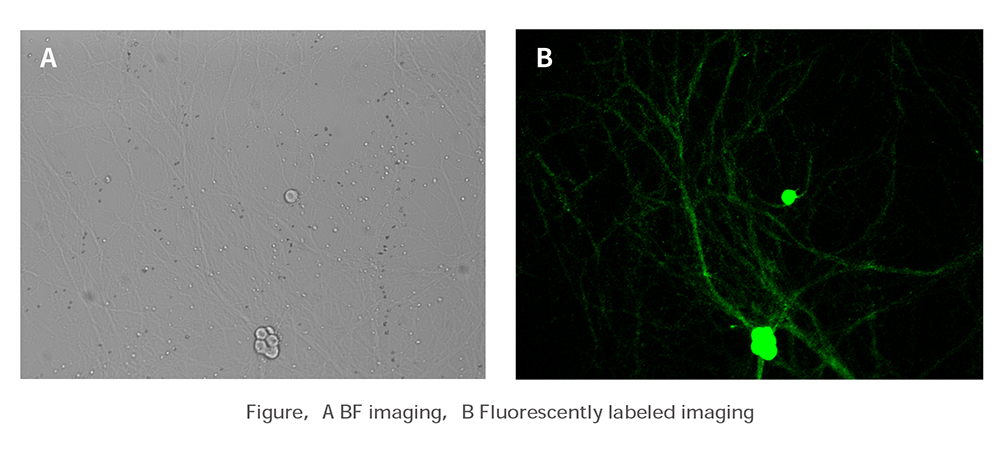
4. Neuronal axon imaging
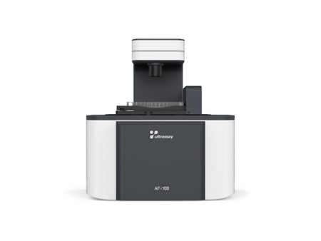
5. Transfection efficiency analysis (AF-100)
6. Organoid imaging
Technical Specification
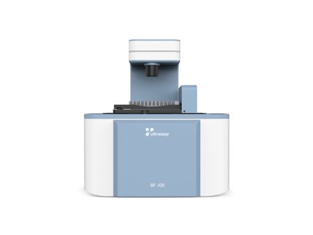
| Mode | UltraView BF-100 | |
| Imaging Channel | Bright field channel | |
| Light Source | White LED | |
| Camera | 12 bit CMOS, 5 megapixels | |
| Objective | 10X standard objective NA 0.25 (4X, 20X are optional) | |
| Imaging Area | 4X: 1.71×1.28mm, 10X: 0.68×0.51mm, 20X: 0.34×0.26mm | |
| Motorized Stage | Automatic travel in X, Y direction, Accuracy: 1μm | |
| Automatic travel in Z direction | ||
| Imaging Mode | 1-field, 4-fields and 9-fields | |
| Data Format | Image: JPEG, TIFF, PNG, Video: AVI, MP4, Data: Csv | |
| Data Output | 2*USB 3.0, RJ45 Port | |
| PC System | Windows 11, CPU i5, 16G RAM, 4TB hard drive, 27-inch display | |
| Comsumables | 6~384wells Microplates, dishes, slides and flasks | |
| Overall Dimension | 328*242*360 mm | |
| Power Supply | 100~240V, 50/60Hz | |
| Working Environment | 5~40°C, 20~95% RH | |
| Net Weight | 9.5 kg | |
Related suggestion
Request for quotation