BRAF mutations are commonly found in malignant tumors such as melanoma, colorectal cancer, thyroid cancer, and lung cancer. Studies have shown that BRAF gene mutation and EGFR, KRAS and other mutations are independent, mutually exclusive and do not appear at the same time [4]. National Comprehensive Cancer Network (NCCN) Clinical Practice Guidelines for Colorectal Cancer (V3.2011) recommends that when patients with metastatic colorectal cancer are treated with targeted drugs such as EGFR inhibitors – Erbitux or panitumumab, KRAS gene mutation detection in tumor tissue should be performed. If KRAS has no mutation, BRAF gene status should be considered. When the patient has V600E mutation, EGFR monoclonal antibody is ineffective. Therefore, understanding the mutation of BRAF gene has become the need to screen EGFR-TKIs and BRAF gene mutation-targeted drugs in clinical targeted drug therapy for the patients who can be benefited.
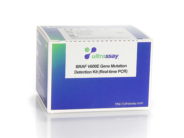
This kit is only used to detect somatic cells mutation of BRAF gene V600E (1799T>A). The test results are for clinical reference only and should not be used as the sole basis for individualized treatment of patients. Clinicians should make comprehensive judgments on the test results based on factors such as the patient’s condition, drug indications, treatment response, and other laboratory test indicators.
UltraDx BRAF V600E Gene Mutation Detection Kit uses PCR enrichment solution, ARMS (Amplification refractory mutation system) technology combined with Taqman probe to detect BRAF gene V600E mutation. The Taqman probe is labeled with FAM that act as a reporter and with BHQ1 which acts as a quencher. When the probe is intact, the fluorescent signal emitted by the reporter is absorbed by the quencher. During the PCR amplification step, the 5′-3′ exonuclease activity of Taq enzyme cleaves and degrades the probe, the reporter fluorophore and the quencher fluorophore are separated, so that the fluorescence monitoring system can receive the fluorescent signal. The PCR enrichment solution reduces the wild-type background sequence through enzyme digestion, and further improves the detection resolution and sensitivity. The characteristics of the ARMS (Amplification Refractory Mutation System) method can specifically recognize the 3′ end of the primer through Taq DNA Polymerase without 3′-5′ exonuclease activity. Taq DNA polymerase is effective at distinguishing between a match and a mismatch at the 3′ end of a PCR primer. When the primer is fully matched, the amplification proceeds with full efficiency. When the 3′ base is mismatched, only ineffective amplification occurs. Therefore, this method can selectively amplify the mutated sequence, and the Taqman probe can be combined with the amplified product to identify the mutated target sequence.
The IC Reference Buffer is used as a quality control for the reagents, DNA quality and the operation itself. Even if the DNA is degraded, the internal control can truly reflect the effective DNA amount of the genes in the system. The detection system contains a certain proportion of dUTP, dNTP and uracil DNA glycosylase (UDG enzyme), this enzyme (also known as uracil-N-glycosylase or UNG enzyme) can interrupt PCR containing dUTP product, thus effectively preventing PCR contamination.
Recommended sample types: Tumor patient’s lesion tissue (papillary thyroid cancer, malignant melanoma, colorectal cancer, lung cancer), including fresh diseased tissue, frozen pathological section, paraffin-embedded pathological tissue or section.
Applied Biosystems™ Real time PCR system 7500, ABI QuantStudio™5 Real-time PCR system, LightCycler® 480 PCR system, Bio-Rad CFX96 real-time PCR instrument. Ultrassay eQ9600 Real Time qPCR System etc.


